


Oct 04, 2006
The neural basis of narrative imagery
The neural basis of narrative imagery: emotion and action.
Prog Brain Res. 2006;156:93-103
Authors: Sabatinelli D, Lang PJ, Bradley MM, Flaisch T
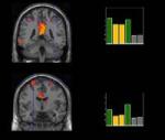
It has been proposed that narrative emotional imagery activates an associative network of stimulus, semantic, and response (procedural) information. In previous research, predicted response components have been demonstrated through psychophysiological methods in peripheral nervous system. Here we investigate central nervous system concomitants of pleasant, neutral, and unpleasant narrative imagery with functional magnetic resonance imaging. Subjects were presented with brief narrative scripts over headphones, and then imagined themselves engaged in the described events. During script perception, auditory association cortex showed enhanced activation during affectively arousing (pleasant and unpleasant), relative to neutral imagery. Structures involved in language processing (left middle frontal gyrus) and spatial navigation (retrosplenium) were also active during script presentation. At the onset of narrative imagery, supplementary motor area, lateral cerebellum, and left inferior frontal gyrus were initiated, showing enhanced signal change during affectively arousing (pleasant and unpleasant), relative to neutral scripts. These data are consistent with a bioinformational model of emotion that considers response mobilization as the measurable output of narrative imagery.
22:43 Posted in Mental practice & mental simulation | Permalink | Comments (0) | Tags: mental practice
Sep 06, 2006
Effects of motor imagery training after spinal cord injury
Effects of motor imagery training after chronic, complete spinal cord injury.
Exp Brain Res. 2006 Aug 31;
Authors: Cramer SC, Orr EL, Cohen MJ, Lacourse MG
Abnormalities in brain motor system function are present following spinal cord injury (SCI) and could reduce effectiveness of restorative interventions. Motor imagery training, which can improve motor behavior and modulate brain function, might address this concern but has not been examined in subjects with SCI. Ten subjects with SCI and complete tetra-/paraplegia plus ten healthy controls underwent assessment before and after 7 days of motor imagery training to tongue and to foot. Motor imagery training significantly improved the behavioral outcome measure, speed of movement, in non-paralyzed muscles. Training was also associated with increased fMRI activation in left putamen, an area associated with motor learning, during attempted right foot movement in both groups, despite foot movements being present in controls and absent in subjects with SCI. This fMRI change was absent in a second healthy control group serially imaged without training. In subjects with SCI, training exaggerated, rather than normalized, baseline derangement of left globus pallidus activation. The current study found that motor imagery training improves motor performance and alters brain function in subjects with complete SCI despite lack of voluntary motor control and peripheral feedback. These effects of motor imagery training on brain function have not been previously described in a neurologically impaired population, and were similar to those found in healthy controls. Motor imagery might be of value as one component of a restorative intervention.
20:45 Posted in Mental practice & mental simulation | Permalink | Comments (0) | Tags: mental practice
Jul 31, 2006
Mapping implied body actions in the human motor system
Mapping implied body actions in the human motor system.
J Neurosci. 2006 Jul 26;26(30):7942-9
Authors: Urgesi C, Moro V, Candidi M, Aglioti SM
The human visual system is highly tuned to perceive actual motion as well as to extrapolate dynamic information from static pictures of objects or creatures captured in the middle of motion. Processing of implied motion activates higher-order visual areas that are also involved in processing biological motion. Imagery and observation of actual movements performed by others engenders selective activation of motor and premotor areas that are part of a mirror-neuron system matching action observation and execution. By using single-pulse transcranial magnetic stimulation, we found that the mere observation of static snapshots of hands suggesting a pincer grip action induced an increase in corticospinal excitability as compared with observation of resting, relaxed hands, or hands suggesting a completed action. This facilitatory effect was specific for the muscle that would be activated during actual execution of the observed action. We found no changes in responsiveness of the tested muscles during observation of nonbiological entities with (e.g., waterfalls) or without (e.g., icefalls) implied motion. Thus, extrapolation of motion information concerning human actions induced a selective activation of the motor system. This indicates that overlapping motor regions are engaged in the visual analysis of physical and implied body actions. The absence of motor evoked potential modulation during observation of end posture stimuli may indicate that the observation-execution matching system is preferentially activated by implied, ongoing but not yet completed actions.
16:45 Posted in Mental practice & mental simulation | Permalink | Comments (0) | Tags: mental practice, motor imagery
Jun 04, 2006
Motor Imagery. A Backdoor to the Motor System After Stroke?
Stroke. 2006 Jun 1;
Authors: Sharma N, Pomeroy VM, Baron JC
BACKGROUND AND PURPOSE: Understanding brain plasticity after stroke is important in developing rehabilitation strategies. Active movement therapies show considerable promise but depend on motor performance, excluding many otherwise eligible patients. Motor imagery is widely used in sport to improve performance, which raises the possibility of applying it both as a rehabilitation method and to access the motor network independently of recovery. Specifically, whether the primary motor cortex (M1), considered a prime target of poststroke rehabilitation, is involved in motor imagery is unresolved. Summary of Review--We review methodological considerations when applying motor imagery to healthy subjects and in patients with stroke, which may disrupt the motor imagery network. We then review firstly the motor imagery training literature focusing on upper-limb recovery, and secondly the functional imaging literature in healthy subjects and in patients with stroke. CONCLUSIONS: The review highlights the difficulty in addressing cognitive screening and compliance in motor imagery studies, particularly with regards to patients with stroke. Despite this, the literature suggests the encouraging effect of motor imagery training on motor recovery after stroke. Based on the available literature in healthy volunteers, robust activation of the nonprimary motor structures, but only weak and inconsistent activation of M1, occurs during motor imagery. In patients with stroke, the cortical activation patterns are essentially unexplored as is the underlying mechanism of motor imagery training. Provided appropriate methodology is implemented, motor imagery may provide a valuable tool to access the motor network and improve outcome after stroke.
22:05 Posted in Mental practice & mental simulation | Permalink | Comments (0) | Tags: mental practice
Feb 28, 2006
Science News: Fitness is good for the brain
From the latest article in Science News Online:
New research suggests that physical exercise encourages healthy brains to function at their optimum levels. Fitness prompts nerve cells to multiply, strengthens their connections, and protects them from harm. Benefits seem to extend to brains and nerves that are diseased or damaged. These findings could suggest new treatments for people with Alzheimer's disease, Parkinson's disease, and spinal cord injuries.
[...]
Preliminary studies indicated that when lab animals exercise, their nerve cells release chemicals called neurotrophic factors. These proteins buffer nerve cells against illness or injury, prompt them to grow and multiply, and strengthen each neuron's connection with other nerve cells.
[...]
Furthermore, memory tests given to 1,740 people over 65 during a 6-year project have linked moderate exercise to reduced risk of dementia. These results were published in the Jan. 17 Annals of Internal Medicine by a Seattle research team.
18:05 Posted in Mental practice & mental simulation | Permalink | Comments (0) | Tags: Positive Technology, mental practice, motor imagery
Dec 22, 2005
Modulation of beta oscillations in the subthalamic area during motor imagery in Parkinson's disease
Brain. 2005 Dec 19;
Authors: Kühn AA, Doyle L, Pogosyan A, Yarrow K, Kupsch A, Schneider GH, Hariz MI, Trottenberg T, Brown P
Activation of the basal ganglia has been shown during the preparation and execution of movement. However, the extent to which the activation during movement is related to efferent processes or feedback-related motor control remains unclear. We used motor imagery (MI), which eliminates peripheral feedback, to further investigate the role of the subthalamic area in the feedforward organization of movement. We recorded local field potential (LPF) activity from the region of the subthalamic nucleus (STN) in eight patients with Parkinson's disease off dopaminergic medication during performance of a warned reaction time task. Patients were instructed to either extend the wrist [motor execution (ME)], to imagine performing the same task without any overt movement (MI), or, in a subgroup, to perform a non-motor visual imagery (VI) task. MI led to event-related desynchronization (ERD) of oscillatory beta activity in the region of the STN in all patients that was similar in frequency, time course and degree to the ERD occurring during ME. The degree of ERD during MI correlated with the ERD in trials of ME and, like ME, was accompanied by a decrease in cortico-STN coherence, so that STN LFP activity during MI was similar to that in ME. The ERD in ME and MI were both significantly larger than the ERD in VI. In contrast, event-related synchronization (ERS) was significantly smaller in trials of MI, and even smaller in trials of VI, than during ME. The data suggest that the activity in the region of the human STN indexed by the ERD during movement is related to the feedforward organization of movement and is relatively independent of peripheral feedback. In contrast, sensorimotor feedback is an important factor in the ERS occurring in the STN area after completion of movement, consistent with a role for this region in trial-to-trial motor learning or the re-establishment of postural set following movements.
22:50 Posted in Mental practice & mental simulation | Permalink | Comments (0) | Tags: Positive Technology, mental practice, motor imagery
Dec 17, 2005
Mental imagery combined with physical practice of approach shots for golf beginners
Percept Mot Skills. 2005 Aug;101(1):203-11
Authors: Brouziyne M, Molinaro C
Recent research on motor skills of golf have pointed to the usefulness of mental imagery. In golf, such training is rarely used as a teaching technique for beginners on the grounds that only top professionals stand to gain from mental imagery. This study tested whether mental imagery combined with physical practice can improve golf performance for the approach shot. 23 volunteer beginners, 8 women and 15 men, M age 23.4 yr. (SD = 3.7), enrolled in the University Physical and Sporting Activities Department, were divided into three groups, using a combination of physical practice of the approach shot plus mental imagery, physical practice only, and a third group engaging in various sporting activities instead of either mental or physical practice of the chip shot. Analysis showed that the beginners' approach shot performance improved most in the group combining physical practice and mental imagery when compared with the group just physically practising the approach shot. It seems mental training can be used effectively to improve performance even with beginners.
12:40 Posted in Mental practice & mental simulation | Permalink | Comments (0) | Tags: Positive Technology, mental practice, motor imagery
Dec 15, 2005
Improvement and generalization of arm motor performance through motor imagery practice
Neuroscience. 2005 Dec 7;
Authors: Gentili R, Papaxanthis C, Pozzo T
This study compares the improvement and generalization of arm motor performance after physical or mental training in a motor task requiring a speed-accuracy tradeoff. During the pre- and post-training sessions, 40 subjects pointed with their right arm as accurately and as fast as possible toward targets placed in the frontal plane. Arm movements were performed in two different workspaces called right and left paths. During the training sessions, which included only the right path, subjects were divided into four training groups (n=10): (i) the physical group, subjects overtly performed the task; (ii) the mental group, subjects imagined themselves performing the task; (iii) the active control group, subjects performed eye movements through the targets, (iv) the passive control group, subjects did not receive any specific training. We recorded movement duration, peak acceleration and electromyographic signals from arm muscles. Our findings showed that after both physical and mental training on the right path (training path), hand movement duration and peak acceleration respectively decreased and increased for this path. However, motor performance improvement was greater after physical compared with mental practice. Interestingly, we also observed a partial learning generalization, namely an enhancement of motor performance for the left path (non-training path). The amount of this generalization was roughly similar for the physical and mental groups. Furthermore, while arm muscle activity progressively increased during the training period for the physical group, the activity of the same muscles for the mental group was unchanged and comparable with that of the rest condition. Control groups did not exhibit any improvement. These findings put forward the idea that mental training facilitates motor learning and allows its partial transfer to nearby workspaces. They further suggest that motor prediction, a common process during both actual and imagined movements, is a fundamental operation for both sensorimotor control and learning.
01:30 Posted in Mental practice & mental simulation | Permalink | Comments (0) | Tags: Positive Technology, mental practice, motor imagery
Nov 30, 2005
Influence of imagined posture and imagery modality on corticospinal excitability
Behav Brain Res. 2005 Nov 25
Authors: Fourkas AD, Ionta S, Aglioti SM
23:25 Posted in Mental practice & mental simulation | Permalink | Comments (0) | Tags: Positive Technology, mental practice, motor imagery
Nov 26, 2005
EMG Activity in Selected Target Muscles During Imagery Rising on Tiptoes in Healthy Adults and Poststroke Hemiparetic Patients
Authors: Dickstein R, Gazit-Grunwald M, Plax M, Dunsky A, Marcovitz E
The authors sought to gain further knowledge about activation of target muscles during imagery engagement in a motor task. Six hemiparetic patients and 9 healthy participants performed 3 real rises on tiptoes and then, after pausing, 3 imagery rises on tiptoes. Metronome beats guided the rate of rises and descents. Electromyographic (EMG) activity from the medial gastrocnemius and the rectus femoris muscles were monitored bilaterally throughout the performance of both tasks. In 3 healthy participants and 3 individuals with hemiparesis, EMG activity was related to the imagery task in at least 1 of the target muscles. Conversely, in the other participants, motor imagery practice was not accompanied by task-related EMG activity in the monitored muscles. In all cases, the increment in activation level during motor imagery practice was very low in comparison with that of real performance. The findings were not unequivocal; therefore, EMG activity may sometimes, but not always, be recorded during motor imagery practice both in healthy individuals and in poststroke hemiparetic participants. Further research is needed to align motor imagery practice with the objectives of motor rehabilitation.
11:55 Posted in Mental practice & mental simulation | Permalink | Comments (0) | Tags: Positive Technology, mental practice, motor imagery
Nov 03, 2005
Mirror therapy for alleviating chronic pain
via Medgadget
McCabe and co-workers from the University of Bath and the Royal National Hospital for Rheumatic Diseases (RNHRD)have published in the journal Clinical Medicine the results of a study, which has investigated the use of mirror as a therapeutic mean to alleviate pain. The treatment consists in asking patients patients suffering from complex regional pain syndrome to carry out routine exercises in front of a mirror.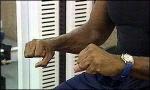
Results showed that more than half experienced pain relief during and after the exercise and further investigations indicated that even greater improvements can be achieved if the tasks are practiced beforehand.
McCabe explain these findings with the ‘cortical’ model of pain. According to this theory, the brain’s image of the body can become faulty, resulting in a mismatch between the brain’s movement control systems and its sensory systems, causing a person to experience pain when they move a particular hand, foot or limb.
Mirror therapy has proven effective also in the treatment of post-stroke hemiplegia as well as in the rehabilitation of "phantom limb" and visual hemineglect.
More to explore
Sathian K, Greenspan AI, Wolf SL. Doing it with mirrors: a case study of a novel approach to neurorehabilitation.
Neurorehabil Neural Repair. 2000;14(1):73-6.
| Ramachandran VS, Altschuler EL, Stone L, Al-Aboudi M, Schwartz E, Siva N. |
Can mirrors alleviate visual hemineglect? Med Hypotheses. 1999 Apr;52(4):303-5.
Altschuler EL, Wisdom SB, Stone L, Foster C, Galasko D, Llewellyn DM, Ramachandran VS.
Rehabilitation of hemiparesis after stroke with a mirror. Lancet. 1999 Jun 12;353(9169):2035-6.
00:45 Posted in Mental practice & mental simulation | Permalink | Comments (0) | Tags: Positive Technology, mental practice, motor imagery
Oct 26, 2005
Theta brainwave triggered training boosts old brains
It has long been known that hippocampus is involved in the learning process. A study published on the Proceedings of the National Academy of Science (USA) has shown that theta brainwave triggered training can enhance learning rate in old rabbits, contrasting the deficit determined by the aging process. This study could have important implications in the development of nonpharmacological treatments for age-related memory deficits.
Nonpharmacological amelioration of age-related learning deficits: The impact of hippocampal  -triggered training
 rhythm, is known to enhance hippocampal plasticity and accelerate learning rate in young subjects, suggesting that manipulations of activity might be used as a means to counteract impairments related to the aging process. Here, young and older rabbits were given eyeblink conditioning trials either when exhibiting hippocampal (+) or regardless of hippocampal activity (yoked control). Although, as expected, older-yoked control animals showed a learning deficit, the older + group learned as fast as young controls, demonstrating that aging deficits, at least in eyeblink classical conditioning, can be overcome by giving trials during episodes of hippocampal activity. The use of several learning criteria showed that the benefits of hippocampal -triggered training in both age groups during the early phase of acquisition, the enhancement persisted in older animals, peaking during later performance. These findings have implications for theories of age-related memory deficits and may contribute to the development of beneficial treatments. depend on different cognitive or motor processes. Whereas there was a benefit of
16:35 Posted in Mental practice & mental simulation | Permalink | Comments (0) | Tags: Positive Technology, mental practice, motor imagery
Oct 22, 2005
Mental rotation in a patient with an implanted electrode grid in the motor cortex
Neuroreport. 2005 Nov 7;16(16):1795-1800
Authors: Tomasino B, Budai R, Mondani M, Skrap M, Rumiati RI
We investigated the effects of cortical stimulation on mental rotation tasks in a patient with an electrode array placed over his left primary motor cortex. The array was implanted to relieve chronic pain resulting from right brachial plexus damage. Tasks involving motor imagery were slowed down by cortical stimulation, whereas those involving visual imagery were not. When the patient performed the motor-imagery task, the interference effect on response times disappeared if the stimulator was switched off. We also probed two of the sites (anterior-lateral and posterior-medial position), and found that stimulation of the more anterior-lateral one consistently disrupted motor imagery.
19:15 Posted in Mental practice & mental simulation | Permalink | Comments (0) | Tags: Positive Technology, mental practice, motor imagery
Sep 08, 2005
Imagery-induced Cortical Excitability Changes in Stroke
Results of this focal transcranial magnetic stimulation study recently published on Cerebral Cortex by a team of Italian neuroscientists suggest that motor imagery could induce a pronounced motor output enhancement in the hemisphere affected by stroke. These findings strongly support the use of motor imagery as a therapeutic mean in post-stroke motor rehabilitation.
Cicinelli P, Marconi B, Zaccagnini M, Pasqualetti P, Filippi MM, Rossini PM.
Cerebral Cortex, 2005 May 4 (Epub ahead of print)
Focal transcranial magnetic stimulation (TMS) was employed in a population of hemiparetic stroke patients in a post-acute stage to map out the abductor digiti minimi (ADM) muscle cortical representation of the affected (AH) and unaffected (UH) hemisphere at rest, during motor imagery and during voluntary contraction. Imagery induced an enhancement of the ADM map area and volume in both hemispheres in a way which partly corrected the abnormal asymmetry between AH and UH motor output seen in rest condition. The voluntary contraction was the task provoking maximal facilitation in the UH, whereas a similar degree of facilitation was obtained during voluntary contraction and motor imagery in the AH. We argued that motor imagery could induce a pronounced motor output enhancement in the hemisphere affected by stroke. Further, we demonstrated that imagery-induced excitability changes were specific for the muscle 'prime mover' for the imagined movement, while no differences were observed with respect to the stroke lesion locations. Present findings demonstrated that motor imagery significantly enhanced the cortical excitability of the hemisphere affected by stroke in a post-acute stage. Further studies are needed to correlate these cortical excitability changes with short-term plasticity therefore prompting motor imagery as a 'cortical reservoir' in post-stroke motor rehabilitation.
12:45 Posted in Mental practice & mental simulation | Permalink | Comments (0) | Tags: Positive Technology, mental practice, motor imagery
Feb 10, 2005
Brain-imaging study on action observation and acquired motor skills
Earlier studies with monkeys revealed that brain cells called “mirror neurons” respond both when we do something, like pick up an object, and when we simply watch someone else do it. It was known that these neurons fire when we perform an action, but it was unexpected that the same cells also fired when we only saw that action being performed. This new study by Calvo-Merino and coll., recently published on the journal “Cerebral Cortex”, goes a step further by showing that such a system operates differently depending on what a person is physically expert at doing.
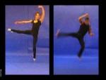
According to the authors of the experiment, this is the first proof that our personal motor repertoire - the things that we have learned to do - changes the way that our brain responds when we see the movement. In particular, these findings suggest that once the brain has learned a skill, it may simulate the skill without even moving, through simple observation: an injured dancer might be able to maintain his skill despite being temporarily unable to move, simply by watching others dance.
These results provide further support to the I-Learning Project clinical rationale, and justify the use of I-Learning technology to better rehabilitate people whose motor skills were damaged by stroke.
Reference: B. Calvo-Merino, D.E. Glaser, J. Grezes, R.E. Passingham and P. Haggard, Action Observation and Acquired Motor Skills: An fMRI Study with Expert Dancers, Cerebral Cortex, Dec. 2004
13:20 Posted in Mental practice & mental simulation | Permalink | Comments (0) | Tags: mental practice, motor imagery